Human heart on a chip could replace animal drug testing
Researchers have created a "heart on a chip" using actual cardiac muscles to help test the effects of heart medication.


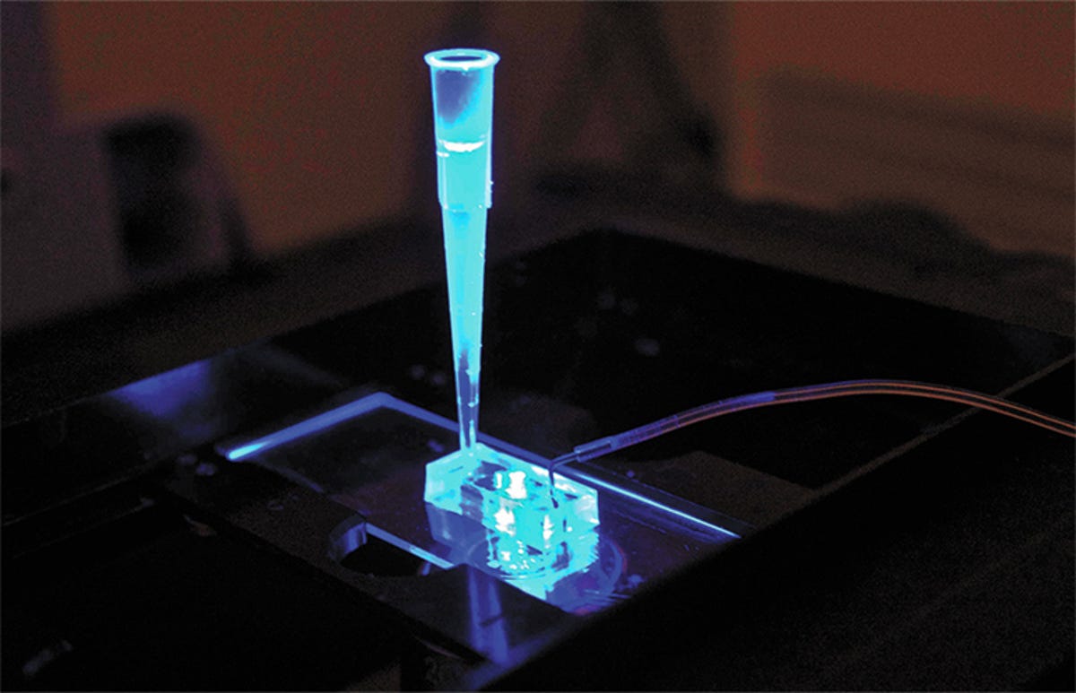
A new device could help make drug testing safer, faster, cheaper -- and eliminate the need for animal testing. It's just an inch long, but inside its silicone body is housed a small piece of cardiac muscle that responds to cardiovascular medications in exactly the same way heart muscle does inside a living human body.
"Ultimately, these chips could replace the use of animals to screen drugs for safety and efficacy," explained Kevin Healy, UC Berkeley professor of engineering, who led the research team that designed the device.
The problems with using animals to test human heart medication aren't merely ethical -- such concerns about lab animals rarely enter scientific discussions. Rather, there are some serious physiological problems -- namely, that drugs designed for humans will not have the same effect on a species that is biologically different from a human.
"These differences often result in inefficient and costly experiments that do not provide accurate answers about the toxicity of a drug in humans," Healy explained.
"It takes about $5 billion on average to develop a drug, and 60 percent of that figure comes from upfront costs in the research and development phase. Using a well-designed model of a human organ could significantly cut the cost and time of bringing a new drug to market."
The chips were created using heart muscle grown in a lab from adult human induced pluripotent stem cells -- stem cells that can be coaxed to grow into many other types of cell. The team then carefully designed the structure to be similar to the geometry and spacing of connective tissue fibre in a living human heart.
Microfluidic channels carved into the silicone on either side of the cell matrix act the same way as blood vessels, mimicking the exchange of nutrients and drugs with human tissue as it would happen in the body.
The cells start beating on their own within 24 hours of being loaded into the chamber at a healthy resting rate of 55 to 80 beats per minute. In order to test the system, the team then administered four well-known cardiovascular drugs -- isoproterenol, E-4031, verapamil and metoprolol. By monitoring the beat rate, the team was able to observe -- and accurately predict -- the chip's response to the drugs. Isoproterenol, for example -- a drug used to treat slow heart rate -- caused the muscle's beat rate to increase from 55 beats per minute to 124 beats per minute half an hour after being administered.
The tissue remains functional for several weeks, allowing each cell matrix to be reused to test multiple drugs.
The use of adult stem cells makes the chip particularly interesting. Researchers could use a patient's own cells to test an individual's responses to various drugs, allowing treatments to be tailored. It could also be modified to model human genetic diseases.
The next step for the team is to determine whether the system could be linked with other organs on a chip to model multi-organ interactions -- for example, a heart on a chip and a liver on a chip.
"Linking heart and liver tissue would allow us to determine whether a drug that initially works fine in the heart might later be metabolised by the liver in a way that would be toxic," Healy said.
The full study, "Human iPSC-based Cardiac Microphysiological System For Drug Screening Applications", can be found online in the journal Scientific Reports.